normal range echocardiography normal values pdf
LV Dimensions Volumes Mass Normal Mild Moderate Severe Normal Mild Moderate Severe LVIDdiastolemm 3756 5761. Arq Bras Cardiol volume 75 nº 2 111-114 2000.
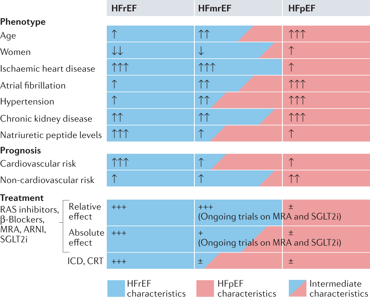
Heart Failure With Mid Range Or Mildly Reduced Ejection Fraction Nature Reviews Cardiology
American Society of Echocardiography Organization of professionals.
. Download full-text PDF Download full-text PDF Read full. In European journal of. When absolute values are.
Lower mean and range than the reference limits. Similarly 3 standard deviations. Recommendations for chamber quantification.
Normal Dog 20 30 circsec. Normal values and thresholds for all heart structures including. 1 Left Ventricle M-mode and 2D a.
Right apical short axis with cursor at level of chordae attachment to papillary muscles. LVEDDBSA mmm2 269 plus or minus 038. AIMS The aim of the Normal Reference Ranges for Echocardiography Study NORRE Study is to obtain a set of normal values for cardiac chamber geometry and function in a large cohort of.
They give the following values for untrained normal individuals the value for trained atheletes are larger. An Update from the American Society of Echocardiography and the European Association of Cardiovascular Imaging 2015 JASE 2015 Jan2811-39e14. Ranges of Left MSAC - Medical Services Advisory CommitteeNormal reference ranges for left and right atrial volume 111 Vertebral Heart Size VHS - Dr.
LVEDDheight mmm 297 plus. Conclusion - The means and estimates of distribution for the measurements of interventricular septum left pos-. This document provides updated normal values for all four cardiac chambers including three- dimensional echocardiography and myocardial deformation when possible on the basis of.
A range of values encompassing 2 standard deviations above and below the population mean includes 964 of all normal subjects. Establishing normal reference values for echocardiography represents a major need in the field of cardiology and the compilation of large databases can be used for this purpose. Manual of Echocardiography.
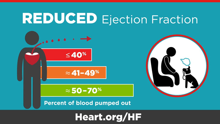
Ejection Fraction Heart Failure Measurement American Heart Association

Swiss Medical Weekly Two Dimensional Transthoracic Echocardiography At Rest For The Diagnosis Screening And Management Of Pulmonary Hypertension

Normal Values Of Right Atrial Size And Function According To Age Sex And Ethnicity Results Of The World Alliance Societies Of Echocardiography Study Journal Of The American Society Of Echocardiography
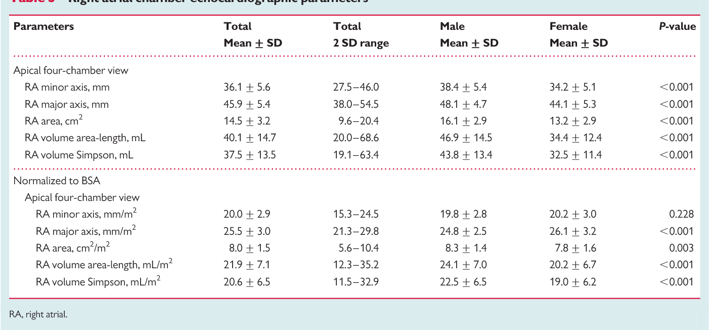
Pdf Echocardiographic Reference Ranges For Normal Cardiac Chamber Size Results From The Norre Study Semantic Scholar
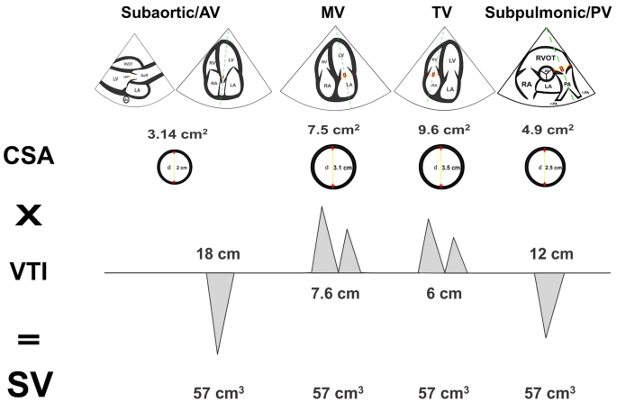
Rationale For Using The Velocity Time Integral And The Minute Distance For Assessing The Stroke Volume And Cardiac Output In Point Of Care Settings The Ultrasound Journal Full Text
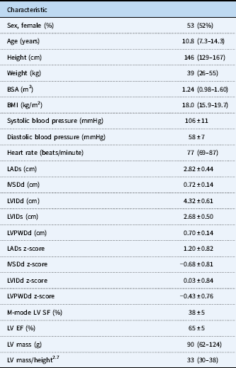
Reference Values For Two Dimensional Myocardial Strain Echocardiography Of The Left Ventricle In Healthy Children Cardiology In The Young Cambridge Core

Normal Values By Age Groups Download Table

American Society Of Echocardiography Iphone Medical App Review

Normal Reference Values Of Echoca Preview Related Info Mendeley

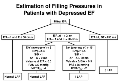
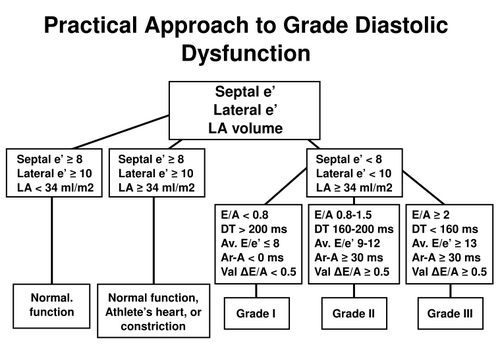
Estimating Left Ventricular Filling Pressure By Echocardiography Sciencedirect
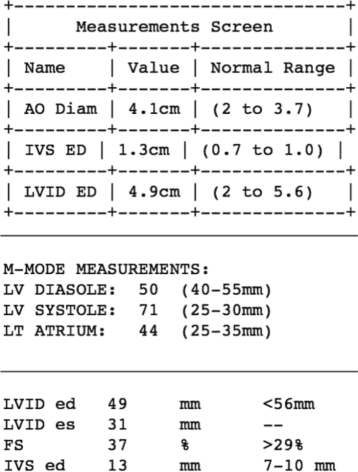
Unlocking Echocardiogram Measurements For Heart Disease Research Through Natural Language Processing Bmc Cardiovascular Disorders Full Text

Echocardiography In The Evaluation Of The Right Heart Usc Journal

Echocardiography The Normal Examination And Echocardiographic Measurements 3rd Edn Bonita Anderson Rrp 159 00 Isbn 13 978 0992322212 Publisher Echotext Australia 362 Pages Prasad 2017 Australasian Journal Of Ultrasound In Medicine Wiley
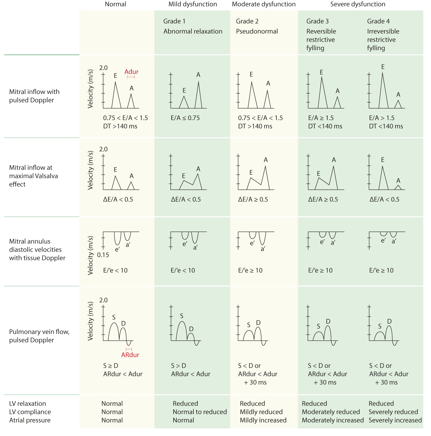
Reference Normal Values For Echocardiography Ecg Echo
Cardiac Time Intervals By Tissue Doppler Imaging M Mode Normal Values And Association With Established Echocardiographic And Invasive Measures Of Systolic And Diastolic Function Plos One

Pdf Normal Echocardiographic Measurements In Indian Adults How Different Are We From The Western Populations A Pilot Study

Left Ventricular Mid Diastolic Wall Thickness Normal Values For Coronary Ct Angiography Radiology Cardiothoracic Imaging
